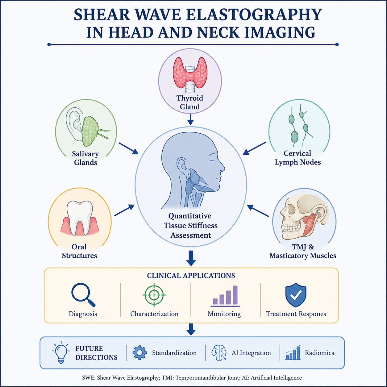
Shear Wave Elastography (SWE) has emerged as a transformative ultrasound-based imaging modality for the quantitative assessment of tissue stiffness, providing valuable insights into the biomechanical properties of normal and diseased tissues. Unlike conventional ultrasonography, which primarily relies on morphological evaluation, SWE enables real-time, reproducible, and relatively operator-independent measurements of tissue elasticity. In recent years, its application has expanded considerably within head and neck imaging, where tissue stiffness serves as a potential biomarker for disease characterization, diagnosis, and treatment monitoring. This narrative review provides a comprehensive overview of the physical principles, quantitative parameters, technical considerations, and clinical applications of SWE in the head and neck region. Current evidence supporting its use in thyroid nodules, salivary gland disorders, cervical lymph node evaluation, oral soft tissue lesions, periodontal assessment, and temporomandibular joint and masticatory muscle disorders is discussed. The integration of SWE with conventional B-mode and Doppler ultrasonography has demonstrated improved diagnostic confidence, enhanced lesion characterization, and reduced reliance on invasive diagnostic procedures in selected clinical scenarios. Despite its promising clinical utility, several challenges remain, including variability in acquisition protocols, machine-dependent elasticity measurements, lack of universally accepted cutoff values, and limited standardization across institutions. Nevertheless, ongoing technological advancements and the growing interest in artificial intelligence, radiomics, and multiparametric ultrasound approaches are expected to further enhance the diagnostic and prognostic capabilities of SWE. Overall, SWE represents a valuable non-invasive adjunct to routine head and neck imaging and holds significant potential as a quantitative biomarker in precision medicine.

 157 (Views)
  87 (Downloads)