
Article
Article ID: 100576
Interaction between Noise Exposure and Phenotypic Age Acceleration in Hearing Loss: An NHANES Study
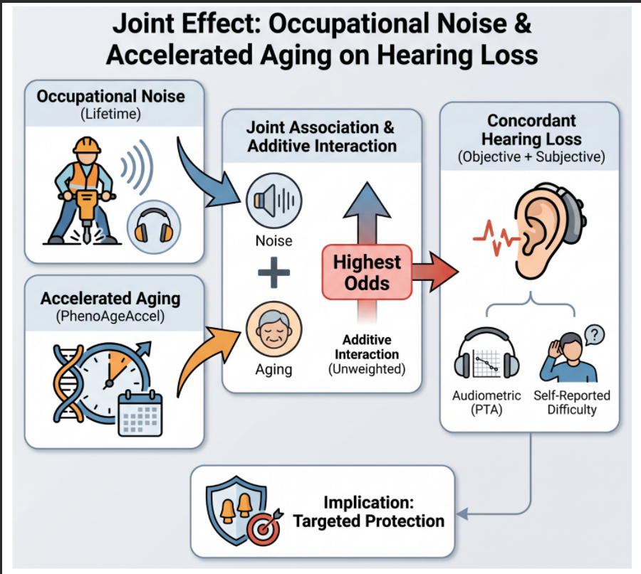
Hearing loss is a growing public health burden, and aging and occupational noise exposure are two major contributors. This cross-sectional study examined whether phenotypic age acceleration interacts with occupational noise exposure in relation to hearing loss. We included 2,014 adults from the National Health and Nutrition Examination Survey 2005–2010, representing 16,596,487 non-institu...