Volume 8 Issue 3 (2018)

Clinical Research

Patients’ Degree of Health Literacy: A Cross-Sectional Survey from Eskisehir, Turkey.

Objectives: The study aimed to evaluate the levels of health literacy amongst patients admitted to the ENT department of Eskisehir Osmangazi University in June 2018.

Methods: 200 patients (comprising 107 males and 93 females), selected at random, consented to the study. They were supplied with the Health Literacy Questionnaire, which was used to evaluate health literacy.

Results: The majority of respondents indicated that they were unsure about whether particular symptoms related to disease or not, lack confidence in being able to read and fully comprehend a text about their disease and have difficulty comprehending and trusting doctors’ advice. They experienced difficulty in weighing advantages and disadvantages of treatment options, finding information relating to symptomatology and particular diseases and being aware when a second opinion would be appropriate. There were deficits apparent in understanding both positive and negative influences of the environment, lifestyle and nutrition. Nutritional advice was poorly comprehended. By contrast, no difficulties were apparent in obtaining medical appointments, making contact with doctors or using medication as directed. Respondents were confident about how to act in an emergency and when to call an ambulance, when to go for checkups and how to locate information on the management and prevention of obesity, hypertension or hypercholesterolaemia.

Conclusion: Health literacy is of prime importance in public health. Bolstering health literacy, especially where levels are low, is necessary. Healthcare staff need to work on being more easily understood in the interests of promoting effective communication across the entire social spectrum.

Read more

Clinical Research

Nasal Complications Related With Cpap Treatment

AIM: In preterm infants who have a tendency for hypoxia, continuous positive airway pressure (CPAP) support is one of the standard treatments being used today. Although newborns are prone to complications, too many complications have not been reported in literature due to CPAP in newborn period. In this study, complications in preterm infants have been reported who had nasal CPAP treatment which were considered to be related to this.

METHOD: Within the scope of the study, 82 babies who were hospitalized in the newborn intensive care unit and had nasal CPAP between September 2014 and September 2017 have been evaluated. During this time period, the newborns that had nasal CPAP treatment have been evaluated in terms of nasal complications in certain intervals.

RESULTS: During this period, complications which are considered to be related to Nasal CPAP have been observed in 6 babies. In 4 newborns, nasal columellar hyperemia related to the use of masks and in 1 newborn, columella necrosis has been seen. In two of the patients, intranasal synechia has been observed. While intranasal synechias were unilateral in 1 patient, it was observed as bilateral multiple nasal synechia in another patient.

CONCLUSION: Continuous positive airway pressure treatment is a lifesaving method when required. However, it may cause certain complications due to pressure and pressured air in the newborn period which is open to all kinds of trauma. It is important to know about these complications and identifying and preventing them in the earliest time possible since the treatment is long and difficult.

Read more

Clinical Research

Evaluation of gustatory functions in patients with laryngopharyngeal reflux

Objective: The aim of the study is to evaluate the gustatory functions in patients with laryngopharyngeal reflux (LPR).

Methods: The study population consisted of the control and the reflux groups. There were 50 patients in each group. Reflux symptom index and reflux symptom scoring system were used in the diagnosis of reflux group. These two groups were subjected to taste test described by Goins et al., and taste test was applied separately to the back, middle and anterior parts of the tongue and the results were compared.

Results: When the demographic characteristics of the groups were compared, there was no statistical difference in terms of sex, age and smoking habit. In our study, we found a statistically significant decrease in the perception of bitterness, sourness and salty taste at the back, mid- dle and 1/3 anterior part of the tongue in the LPR group when com- pared to the control group. There was no significant difference between groups in terms of sweet taste.

Conclusion: While laryngopharyngeal reflux disease affect negatively the salty, bitter and sour taste functions, we found the sweet taste as the most resistant taste to LPR. Histopathological examination in animal experiments may be useful in order to prove the hypothesis concerning loss of taste caused by LPR.

Read more

Clinical Research

Investigating CPAP Compliance in Patients with Obstructive Sleep Apnea

Objective: Our aim in this study is to investigate, in patients who are advised to use CPAP, the rate of usage, differences between the patients who use the device and those who don’t; and the factors affecting the compliance rates.

Methodology: A telephone survey was applied to the patients who underwent polysomnography, between the years 2010 -2015 and were thereafter advised to use CPAP as the treatment method.

Results: Total number of patients included in the study was 108. Of them, 77 (71%) was male and 31 (29%) was female. The mean age was 50 (±11). There were 47 (43,5%) patients with comorbidities. Of 108 patients, 63 (58%) reported using CPAP at the time of the survey and 45 (42%) reported having abandoned the treatment, 62% (n: 28) of this 45 patients reported that they ceased the CPAP use within the first week of their treatment. Of 63 patients using CPAP, 3 (5%) was found noncompliant while 60 (95%) was found compliant.

Conclusion: In our study, rate of CPAP use was found to be 55%. The difference between the mean ages of CPAP users and non-users was found statistically significant (p: 0,008). the results of our study suggests that BMI is not a factor that affects the rate of CPAP use. In our study, contrary to what would be expected, the mean CPAP pressure of the patients using the device were found to be higher than that of non-users.

Read more

Clinical Research

Evaluation Of Autologous Serum Skin Testing Results In Patients With Seasonal Allergic Rhinitis

Objective: Autologous serum skin testing is a practical test for basophil histamine release which is used in the diagnosis of chronic autoimmune urticaria. Although the exact autoimmune pathogenesis of allergic diseases of the upper respiratory tract remains a matter of debate, recent studies have reported positive results for autologous serum skin test in both allergic and non-allergic rhinitis, asthma sufferers, and even in healthy controls. In this study, we aimed to evaluate positive results from autologous serum testing in patients with allergic rhinitis and to compare the results with those from healthy controls.

Methods The study included 76 seasonal allergic rhinitis patients and 51 healthy volunteers aged between 18-65 years who underwent autologous serum skin testing. A chi-square test was used for comparisons between the two groups.

Results Autologous serum testing proved positive in 29 of the patients with allergic rhinitis and in 10 of the healthy controls. The number of positive results from autologous serum testing was found to be significantly higher in patients with allergic rhinitis than in the control group.

Conclusion Autologous serum skin testing may have benefits beyond merely detecting chronic urticaria. In the present study, the number of allergic rhinitis patients who demonstrated autologous serum skin testing positivity was significantly higher than the healthy controls. The findings of the present study may contribute further studies that investigate common pathological mechanisms of allergic rhinitis and chronic idiopathic urticaria patients who show positivity to autologous serum skin testing.

Read more

Clinical Research

Comparison Of Post-Tonsillectomy Morbidity In Cases Of Sleep Apnoea Where Surgery Employed Either Cold Knife Or Coblation Techniques

Objective: To compare the snare and coblation methods in terms of post-tonsillectomy morbidity for patients with obstructive sleep apnoea.

Method: The study involved 49 cases. Twenty-one patients were operated on using the snare method, whilst the remaining 28 patients underwent coblation tonsillectomy. The amount of intraoperative bleeding was recorded. The pain level at the 6th and 12th postoperative hour was assessed. On postoperative days 1, 3 and 7, the patients were assessed through VAS (Visual analogue scale) and Wong-Baker Faces Pain Rating Scale (WBFPRS). The two groups were then compared. The Tonsillar Fossa Wound Healing Score (TFWHS) assessment was also undertaken to assess wound healing rates on postoperative days 1, 3, 5, 7 and 10.

Results: The patients who underwent coblation had significantly lower amounts of bleeding compared to those whose operations employed the snare method (p=0.046). Those patients who underwent tonsillectomy using the snare method had significantly higher 6th hour VAS and WBFPRS scores (p=0.011, p=0.005) than the other group. In contrast, VAS and WBFPRS scores at the 12th hour postop and on days 1, 2, 3 and 7 were similar between the groups. TFWHS for those who underwent coblation were significantly higher on days 1 and 3 (p=0.007; p=0.008). However these scores were similar on days 5, 7, and 10.

Conclusion: The research indicates that the amount of intraoperative bleeding in cases undergoing coblation is significantly lower. Lower pain in the early postoperative period, resulting in earlier oral intake and better recovery scores for the tonsillary fossa seem to be the advantages of coblation as opposed to the snare method.

Read more

Clinical Research

Postural Mechanisms That Maintain Airway Adequacy In Obstructive Sleep Apnoea As Determined By Magnetic Resonance Imaging

Objective: This magnetic resonance imaging (MRI) study aimed to explored associations between neutral head posture, size, and shape of the pharyngeal airway and the severity of obstructive sleep apnoea (OSA).

Methods: A retrospective methodology was used to study 90 male patients who had already undergone overnight polysomnography and cervical MRI. 60 cases of OSA were compared with a control group of 30 mild OSA (or straightforward snoring) cases in terms of MRI, with the aim to examine how neutral head posture, the pharynx and the adjacent anatomical structures interact. MRI was performed in all cases with the patient supine and head held in neutral position. Measurements were taken of the Craniocervical extension (CCE) and epiglot angle, length of the tongue root, distance between the hyoid and the plane of the mandible (MP-H distance), and the diameter of the pharyngeal airway at seven points were measured.

Results: Differences in shape were more perceptible at the caudal levels. In the OSA group, the shapes were more oblique. The retroglossal level was where the largest shape difference was apparent. After adjusting for body mass index and age, neutral head posture was correlated with OSA severity. There was a correlation between CCE and lengthening of the tongue root, MP-H distance, epiglot angle, and the two most caudal airway areas.

Conclusions: Overall, increased length of the root of the tongue, MP-H distance, and epiglot angle are associated with CCE in OSA patients and resulted in a larger and more oblique airway in the majority of caudal planes. Such an alteration may be viewed as an adaptation in posture designed to keep the airway sufficiently open in patients suffering from OSA.

Read more

Clinical Research

Normalisation Values For The Resonant Frequency Of The Middle Ear In Rats

Objective: In earlier studies, the resonant frequency (RF) of the middle ear has been investigated in a number of different animals. However, no study has so far specifically addressed the measurement of RF in rats. With this in mind, in our study, multifrequency tympanometry (MFT) was performed on rats and RF value measurements were taken. The aim was to ascertain the normal values for rats used in our university experiments and to provide guidance for further research to be undertaken in this area.

Materials and Methods: For the study, 16 male and 16 female adult Sprague Downey rats (32 animals, 64 ears) aged 10 months old and in healthy condition were used. MFT at 226Hz frequency and above was performed on all the rats after they had been anaesthetised. In the first instance tympanograms were recorded using the 226Hz probe tone, following which MFT measurements were performed. RF values for the middle ear were determined in the range 200-2000Hz.

Results: For male rats, the mean RF value was 426.56 (± 193.01 Hz), whilst for female rats the mean was 496.88 (± 132.55) Hz. When the values obtained for the male and female rats were compared, no statistically significant difference was observed between the two groups (p=0.061). Accordingly, the mean RF value for the rats as a whole (obtained for 64 ears) was calculated to be 461.7 (± 168.02) Hz.

Conclusion: In this research, RF values for the middle ear of rats has been demonstrated to be measurable. However, further research is needed into the relationship between RF and the characteristics of the middle ear in rats.

Read more

Clinical Research

Evaluation of Anatomical Variations on Paranasal Sinus CT

Objective: The purpose of this study was to analyze and assess the frequency of anatomical variants of the paranasal sinuses as detected by multidetector computed tomography (MDCT) scanning.

Methods: 225 patients (122 males, 103 females) in total, with a median age of 28 (range: 15-77) years were included in this retrospective study. Paranasal computed tomography was conducted in all cases. The scans were reviewed to discover the incidence of nasal septal deviation (NSD), variations of the nasal turbinates, ethmoidal air cells, paranasal sinus pneumatization, accessory pneumatization of the paranasal complex, variations in the sphenoid sinus-related structures and the optic nerve and Vidian canal classification type.

Results: NSD was present in 124 (55.1%) of individuals, superior concha pneumatization in 30 (13.3%), middle concha pneumatization in 100 (44.4%), Agger nasi cells (ANC) in 192 (85.3%), Haller cells in 47 (20.9%), Onodi cells in 44 (19.6%), maxillary sinus hypoplasia in 6 (2.6%), frontal sinus hypoplasia in 29 (12.9%), frontal sinus aplasia in 7 (3.1%), sphenoid sinus hypoplasia in 7 (3.1%), uncinate process pneumatization in 12 (5.3%), anterior clinoid process (ACP) pneumatization in 60 (26.7%) and pterygoid process (PP) pneumatization in 75 (33.3%). Optic nerve Type 1 was found in 122 (54.2%) individuals, Type 2 in 29 (12.9%), Type 3 in 36 (16%) and Type 4 in 38 (16.9%). Vidian canal Type 1 was observed in 37 (16.4%), Type 2 in 95 (42.2%) and Type 3 in 93 (41.3%) individuals.

Conclusion: The analysis of paranasal CT is crucial in the diagnosis of miscellaneous diseases of the paranasal sinuses. Anatomical variations in the sinuses are highly prevalent, so otorhinolaryngologists should be aware of these variations to allow them to reach precise diagnoses and avoid surgical complications.

Read more

Clinical Research

Diagnostic Performance of Diffusion-Weighted Magnetic Resonance Imaging in Patients with Cholesteatoma

Objective: Our aim was to evaluate the diagnostic performance of Magnetic Resonance Imaging (MRI) with diffusion-weighted images in patients with cholesteatoma.

Methods: We compared the preoperative MRI findings and intraoperative microscopic examination findings in 54 patients who were operated on due to a pre-diagnosis of chronic otitis media with cholesteatoma, according to preoperative microscopic ear examination, temporal bone computed tomography (CT) and ear MRI.

Results: Fifty-four patients (18 female and 36 male) were enrolled in this study. The mean age was 36.8 ± 17.3 (range: 6-67). Thirty-one patients had primary surgery, whereas 23 patients had revision surgery to the affected ear (for 19 patients, the second, for 3, the third, and for 1, the 5th operation).We found that the sensitivity of pre-operative MRI for detecting cholesteatoma was 97.7%, the specificity was 77.8%, and the diagnostic accuracy rate was 94.4%. The rate of false negatives in MRI with diffusion-weighted images was found to be 4% in primary cases, whereas the false negativity rate with this technique was found to be 0% for revision cases.

Conclusion: We concluded that ear MRI examination with diffusion-weighted images is a significant diagnostic tool, to be used alongside preoperative history and physical examination in deciding on whether to operate, especially in patients for whom revision surgery of the ear (due to recurrent, residual, iatrogenic cholesteatoma, or squamous epithelium) is planned.

Read more

Clinical Research

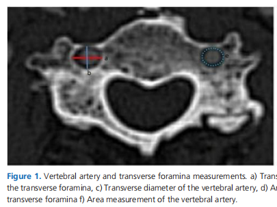
Anatomical Considerations: The Relationship Between The Vertebral Artery And Transverse Foramina At Cervical Vertebrae 1 To 6 In Patients With Vertigo

Objective: In this study, we aimed to investigate the relationship between the size of the vertebral artery and that of the transverse foramina at the C1 to C6 vertebral level in patients suffering from vertigo thought to be related to vertebrobasilar insufficiency (VBI).

Materials and Methods: In this retrospective study, two groups were compared in terms of cervical computed tomography (CT): 22 adult patients with vertigo; and a control group consisting of 23 healthy adult indivıduals. Measurement of the vertebral artery and transverse foramina (i.e. sagittal and transverse dimensions, plus area) were performed bilaterally at levels C (cervical vertebra) 1 to C6.

Results: For the cases group, at C6 level the right vertebral artery area, sagittal and transverse diameter were larger than in the control group at the level of statistical significance. At C1 level, the area of the right transverse foramina in the cases group was significantly higher than in the control group. At levels C1 to C5, the values obtained for vertebral artery area were positively correlated with the values for the ipsilateral transverse foramina (transverse foramen sagittal and transverse dimensions plus areas). Where the sagittal or transverse dimensions of the transverse foramina, or the area of the transverse foramina were found to be decreased, vertebral artery areas were also observed to have decreased at C1 to C5 levels.

Conclusion: We concluded that a decrease in diameter of the bony structures or transverse foramina may cause a decrease in the cross-sectional area of the vertebral artery on the ipsilateral side. Since it is only the left vertebral artery which is dominant for cerebral blood flow, any compensatory increase in right vertebral artery area cannot offset decreased cerebral blood flow. Decreased blood flow (i.e. VBI) on the left side may play a role in the development of vertigo.

Read more