Volume 12 Issue 3 (2022)

Original Article

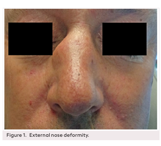
Effect of Total Intravenous Anesthesia and Inhalation Anesthesia on Edema and Ecchymosis in Rhinoplasty: A Prospective Randomized Clinical Trial

Background: Postoperative edema and ecchymosis are frequently observed in rhinoplasty patients. Total intravenous anesthesia and inhalation anesthesia are 2 of the common types of general anesthesia used for rhinoplasty surgery and affect circulation and coagulation. The aim of this study was to investigate the edema and ecchymosis effects of total intravenous anesthesia and inhalation anesthesia in patients undergoing rhinoplasty.

Methods: A total of 52 patients undergoing rhinoplasty surgery were enrolled. The patients were divided into 2 groups: total intravenous anesthesia (group T, n=26) and inhalation anesthesia (group S, n=26). All patients were photographed by a blinded observer on postoperative days 1, 2, and 7 and were evaluated on a scale ranging from 0 to 4 by 3 independent observers for periorbital edema and ecchymosis. Blood samples were taken from the patients preoperatively and at the second postoperative hour. At the end of the study, edema and ecchymosis of the patients, coagulation, and blood parameters were compared according to the anesthesia type.

Results: When rhinoplasty patients in group T and group S were evaluated according to edema and ecchymosis on postoperative days 1, 2, and 7, no statistically significant difference was found (P > .05). There was no difference between the groups in terms of coagulation and blood parameters (P > .05).

Conclusion: No difference between total intravenous anesthesia or inhalation anesthesia according to edema and ecchymosis can be seen in the postoperative period in rhinoplasty patients.

Read more

Original Article

Evaluation of Superficial Parotidectomy Videos on Youtube in Terms of Surgical Education

Background: This study examined the videos of superficial parotidectomy on Youtube and aimed to evaluate whether these videos meet the basic educational steps with laparoscopic surgery video educational guidelines.

Methods: We analyzed the results of a Youtube search with the keywords “superficial parotidectomy surgery, superficial parotidectomy, parotidectomy” until February 2020. After the exclusion criteria, we evaluated the remaining 38 videos with parameters such as the total number of views, subscribers, likes and dislikes, comments, etc. We also analyzed the videos with the laparoscopic surgery video educational guidelines’ video quality assessment tool.

Results: Of the 38 evaluated videos, 10 were high quality (26.3%) and 28 were low quality (73.7%). No statistically significant difference was observed between the study groups in the rates of music, the number of subscribers, comments, video age (days), likes/subscribers, likes/views, views/subscribers, and likes (P> .05 for all comparisons). On the other hand, the number of views, the number of likes, (likes × views)/100 ratios, (likes−dislikes)×100, likes+dislikes, likes−dislikes, and Video Power Index are significantly higher for high-quality videos (for all comparisons, P < .05). As expected, all laparoscopic surgery video educational guideline scores were significantly higher for all parameters in the high-quality video group, while no significant difference was observed for the sixth item (P=.386).

Conclusion: We found that popular YouTube videos about superficial parotidectomy surgeries were significantly lacking in information regarding case presentation, treatment options, intraoperative and postoperative complications, and the healing process. We believe that videos used as a source of information should be recorded by more qualified professionals and that the content should be presented with these missing features.

Read more

Original Article

Histopathological Examination of Cyanoacrylate Usage in Cartilage Graft Fixation

Background: The aim of this study is to investigate the histopathological effects of 2-octyl-cyanoacrylate used in facial plastic operations that are applied for the fixation of cartilage graft types and the graft stabilization effect.

Methods: In the study, 16 New Zealand-type rabbits weighing between 2250 g and 2500 g, 3-4 months old, were used. Two groups were formed as the study and control groups. Those using 2-octyl-cyanoacrylate for cartilage graft fixation were defined as the study group. Those using suturing were defined as the control group. The operation was planned with cartilage grafts taken from the ears of each rabbit and the study group on the left of the sagittal suture and the control group on the right.

Results: Acute inflammation findings were found in 19 (39.6%) of the study group and 6 (14%) of the control group. Acute inflammation levels were found to be higher in the study group than in the control group (P=.006). Mild inflammation was found in 20 (41.7%) of the study group, moderate inflammation in 7 (14.6%), and severe inflammation in 2 (4.2%). Mild inflammation was detected in 10 (23.3%) of the control group. Chronic inflammation levels were higher in the study group than in the control group. Foreign body reaction was detected in 28 (58.3%) of the study group and 14 (32.6%) of the control group. Foreign body reaction levels were found to be higher in the study group than in the control group.

Conclusion: We think that the use of 2-octyl-cyanoacrylate in cartilage graft fixation is not correct as it may increase the risk of postoperative complications.

Read more

Historic Communications

First Electrical Stimulations of the Ear with Voltaic Pile (Letter by Professor Volta to Professor Brugnatelli, “Above the Application of Electricity to Deaf-Mutes from Birth”)

In the late 18th century, several European scientists started describing the clinical effects of different types of electrical stimulation and tried to find new treatments for medical conditions, but only in the very early years of the 19th century, first attempts to cure deafness began. The letter written between 2 famous Italian scientists in the second half of 1802, by Professor A. Volta to Professor L.V. Brugnatelli, “Above the application of electricity to deaf-mutes from birth,” is one of the most detailed examples of how the experiments were conducted applying the voltaic pile as a cure for deafness. In order to gain a better understanding of the context in which the scientists of the time experimented with “medical electricity” on the ear, some of the experiments conducted by European scientists will be described in this study, including those conducted by J.J.A. Sprenger, F.L. Augustin, C.H. Wolke, C.J.C. Grapengiesser, and E.A. Eschke.

Read more

Case Report

Nasal Gout: A Rare Pathology

Gout is the most frequent cause of inflammatory arthritis. However, it may present atypical deposit sites for monosodium urate crystals, making its diagnosis difficult, particularly if there is no previous history of hyperuricemia. The aim of this study is to present a rare form of presentation of a common pathology such as gout with tophaceous gout involving the nasal dorsum as the first presentation. We report a 47-yearold male who presented at our clinic due to nasal deformity with progressive growth and a painful lesion located in the right nasal ridge. Due to the absence of other complaints or associated pathologies, and regarding CT evaluation which confirm a lesion on the right side of the nasal pyramid, with apparently well-defined limits, with an anteroposterior axis of approximately 10 mm, no diagnosis was possible without histologic findings. The patient underwent surgery to excise the lesion and the histological evaluation of the surgical specimen revealed a gouty tuft. Due to the aesthetic deformity, grafting was performed with cartilage and temporal fascia. An analytical evaluation confirmed hyperuricemia at the same time that another gouty lesion appeared on the left hand. He was referred to a rheumatology consultation and started medication with allopurinol. Thus this case draws our attention to the fact that common pathologies can have very atypical presentations.

Read more

Case Report

An Extraordinary Case of Maxillofacial Trauma: Injury by Harpoon Shot

This case report presents a 24-year-old patient who accidentally received maxillofacial trauma with a speargun. One of the harpoons on the right and the other on the left had penetrated the submandibular region, and the floor of the mouth created lacerations on both sides of the tongue and extended into the nasal cavity by penetrating the hard palate bilaterally. In the maxillofacial computed tomography of the patient, the tip of the harpoon on the right ended in the right nasal cavity; however, it was seen that the tip of the speargun on the left side penetrated the frontal sinus on the left side. Both orbits and intracranial areas were regular. The patient was taken to an emergency operation. Tracheotomy was performed under local anesthesia to ensure airway safety. The harpoon on the left was removed from the body in the direction of the entrance with the help of forceps. While the harpoon on the right was removed in the direction of the entrance, the harpoon was removed by exploring the submandibular region due to the hook opening at the mouth’s floor. The patient, who had no complications in the postoperative follow-up, was discharged on the ninth postoperative day.

Read more